Dental X-rays
An examination can only assess what is visible. X-rays cover the rest — between the teeth, inside them, and in the bone underneath. We use digital imaging at our Vienna office, minutes from Tysons Corner, and we take images when they will tell us something rather than on a fixed timer.
Why dental X-rays are necessary
Roughly speaking, an exam sees three of the five surfaces on a tooth. The two it cannot see are the ones facing the neighbouring teeth, and those are exactly where adult decay tends to begin. A cavity can be well established between two molars while both look perfectly sound from above — and it will not hurt until it is deep, because the outer layers of a tooth have no nerve supply.
Everything below the gum line is invisible too. That includes the bone that holds your teeth in place, the roots, the tips of the roots where infection collects, wisdom teeth that have not come through yet, and cysts or lesions that produce no symptoms at all until they are large. None of that can be assessed by looking, and none of it can be assessed by asking how you feel.
The purpose of imaging is not thoroughness for its own sake. It is to find things while they are still cheap and simple to deal with — a filling rather than a root canal, a watch-and-wait rather than an extraction.
The types of dental X-ray, and what each one is for
Different images answer different questions, which is why you will not have all of them at every visit.
Bitewing
You bite gently on a tab and the image captures the upper and lower back teeth together. This is the workhorse: it detects cavities between teeth and shows the bone level around them. For most adults these come round about once a year.
Periapical
A view of one or two teeth in full — crown, root, and the bone at the root tip. This is what we take when a specific tooth is aching, when infection is suspected, or when planning root canal treatment, an extraction, or an implant.
Digital panoramic
A single image giving a full view of the jaw, the alveolar bone, and the roots of the teeth. Taken every three to five years, and the one that shows wisdom teeth, the jaw joints, sinuses, and anything unexpected sitting in the bone.
A first visit here usually includes a comprehensive set, because we are establishing a baseline rather than checking against one. After that, imaging is driven by what the previous images showed and how your mouth is behaving.
The scan that is not an X-ray at all
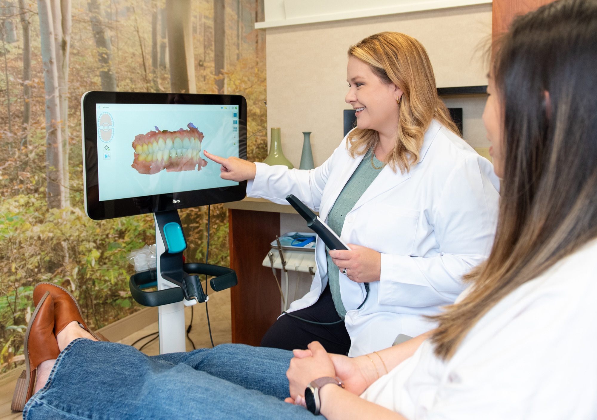
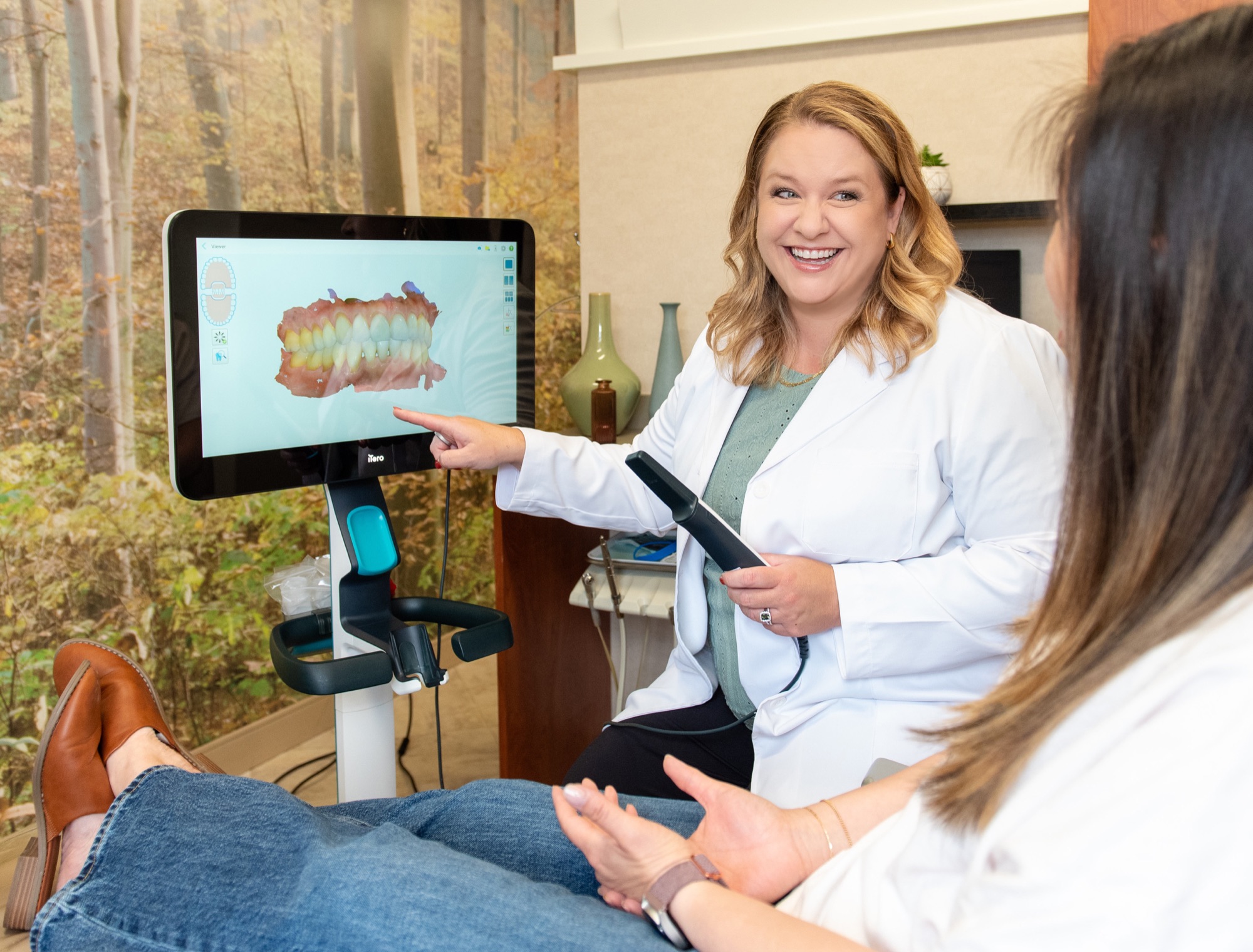
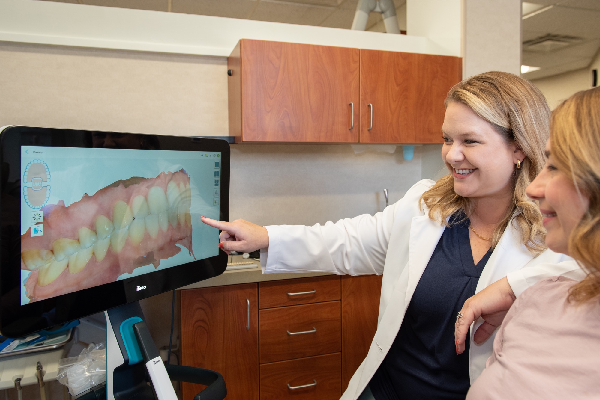
Alongside imaging we offer the iTero Health Scan, and it is worth being clear that it is a different thing. A wand photographs the surfaces of your teeth and gums and assembles a three-dimensional model on the screen beside you, in three to five minutes, using no radiation whatever. It cannot see inside a tooth or into bone, so it does not replace an X-ray. What it does well is show surfaces and shape — wear, recession, crowding, how the bite meets — and it gives us a dated record to compare against in a couple of years. It is free to every patient here.
How much radiation is actually involved
Dental X-rays yield relatively low radiation compared with other sources, and the useful way to think about it is by comparison rather than in the abstract. A single dental X-ray is about 0.2 microsieverts. A flight from New York to Los Angeles exposes you to roughly 10 — some fifty times more, and nobody weighs that up before booking a ticket.
Digital sensors are part of why the figure is low; they need far less exposure than the film they replaced, and the image appears immediately with no chemical processing, so a retake is rare. We use a lead apron and thyroid collar, we collimate the beam to the area of interest, and we take the smallest number of images that will answer the question in front of us.
If you are pregnant, tell us. Routine imaging is generally deferred as a precaution, though a necessary X-ray during pregnancy is considered safe with shielding — an untreated infection carries its own risks, and that trade-off is a conversation to have rather than a rule to apply. Children need imaging less often than adults, and we keep it to what is genuinely needed.
What radiographs mean to you as a patient
Two things, mainly. The first is that a diagnosis stops being an opinion you have to trust. The image is on the screen, Dr. Lokitis can point at the dark shadow between two molars, and you can see the same thing she is seeing. That matters most when the recommendation is to do nothing yet — watching a small lesion is much easier to accept when you have looked at it.
The second is that your images become a record. Bone levels, root shapes, old restorations, that slightly odd spot on a lower premolar: all of it is dated and kept. Comparing this year against three years ago is often more informative than either image alone, because dentistry is mostly about rate of change. Something static for a decade is a different problem from something that moved in eighteen months.
Your images belong to you. If you move away or change practice, ask and we will send them on. If you are joining us from another dentist, tell us before your appointment and we will try to request your recent images rather than repeat them. Imaging supports the rest of preventive care — cleanings and exams, oral cancer screening, and general dentistry — rather than standing on its own. Questions are welcome: contact us or call (703) 893-1603.
Frequently Asked Questions
Are dental X-rays safe? expand_more
Dental X-rays yield relatively low radiation compared with other everyday sources. A single dental X-ray is around 0.2 microsieverts; a flight from New York to Los Angeles is roughly 10, about fifty times more. Digital sensors need less exposure than film did, and we use a lead apron and thyroid collar and take only the images that will answer the question in front of us.
How often will I need X-rays? expand_more
It depends on your mouth rather than the calendar. For most adults bitewings come round about once a year, more often if you are prone to decay or have active gum disease, and a digital panoramic image every three to five years. A first visit usually includes a comprehensive set because we are establishing a baseline.
Can I have X-rays while I am pregnant? expand_more
Tell us if you are pregnant or think you might be. Routine imaging is generally postponed as a precaution. Where an X-ray is genuinely needed — a suspected infection, for instance — it is considered safe with proper shielding, and leaving an infection untreated carries its own risks. It is a conversation rather than a blanket rule.
Do children need dental X-rays? expand_more
Sometimes, and less often than adults. Images can show decay between baby teeth, whether adult teeth are forming and coming through in the right places, and how much room there is for them. We keep children's imaging to what is genuinely useful, and we explain what we are doing before we do it.
Can I bring X-rays from my previous dentist? expand_more
Yes, and please do. Tell us before your appointment and we will try to request them, which may save you a retake. Your images belong to you — if you leave us, ask and we will forward them on.
Is the iTero scan a type of X-ray? expand_more
No. The iTero scan is optical: a wand photographs the surfaces of your teeth and gums and builds a 3D model, with no radiation at all. It shows shape and surface — wear, recession, crowding, how the bite meets — but it cannot see inside a tooth or into bone, so it complements X-rays rather than replacing them. It is free to every patient here.
Why do I need a panoramic X-ray if I have just had bitewings? expand_more
They show different things. Bitewings are detailed close views of the back teeth and the bone between them. A panoramic image is a single wide view of the whole jaw, including wisdom teeth, the jaw joints, the sinuses, and areas of bone that bitewings simply do not reach. That is why the panoramic comes round every three to five years rather than annually.
Ready to Get Started?
Contact our Vienna office to schedule your appointment with Dr. Julia Lokitis.